
 Before the advent of microsurgery in the mid-1960s,
neurosurgeons relied on anatomical drawings little changed from the time
of Leonardo daVinci to guide them through a part of the body where even
the slightest misstep can result in death or devastating debilitation for
the patient.
Before the advent of microsurgery in the mid-1960s,
neurosurgeons relied on anatomical drawings little changed from the time
of Leonardo daVinci to guide them through a part of the body where even
the slightest misstep can result in death or devastating debilitation for
the patient.
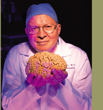
Into this environment came Albert Rhoton
Jr., a young man only a few years removed from a rustic cabin and two-room
schoolhouse in rural eastern Kentucky. Rhoton quickly realized the microscope’s
potential, not only to revolutionize surgery itself but to take brain anatomy
to a new level of detail.
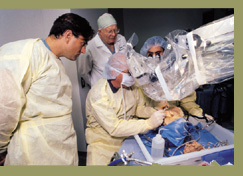
For the first time, the surgical microscope
enabled anatomists to map the brain’s interior landscape using more
than the naked eye. Magnification brought into focus previously invisible
blood vessels branching off and draining from cerebral arteries and previously
unknown connections among vital centers of the brain.
"Knowledge of brain anatomy as studied
under magnification remains the most crucial aspect of the vast refinements
I’ve witnessed in neurosurgical practice,” says Rhoton. “As
surgeons, we need to determine the most gentle and least traumatic, tissue-preserving
approach we can take to reach a specific site to perform corrective surgery,
and anatomical studies through the microscope have helped us do that.”
Now in the 36th year of an illustrious career, Rhoton is regarded by brain
surgeons the world over as the father of microscopic neurosurgery.
Last fall, in recognition of his work, a textbook-sized collection of Rhoton’s
updated papers and illustrations on the brain’s posterior cranial fossa
region was published as part of the international journal Neurosurgery.
The unique “millennium supplement” was designed to serve as a
guide to surgeons operating within this critical region of the brain, containing
nerves that help to control such vital functions as breathing, blood pressure,
balance, consciousness, hearing and vision.
“Anatomy to the surgeon is like the sun for our planet; it gives us
the life-sustaining knowledge to traverse the intricate pathways throughout
the brain,” Arizona neurosurgeon Robert Spetzler wrote in a preface
to the Neurosurgery supplement. “No single neuroanatomist can lay greater
claim to expanding this knowledge for neurosurgeons than Dr. Albert L. Rhoton
Jr.”
With typical humility, Rhoton expresses amazement at a career track that
has taken him from a disadvantaged childhood that began with a midwife assisting
his birth in exchange for a sack of corn to a position as one of the world’s
preeminent neurosurgeons.
Rhoton continues to view the mystery and wonder of the brain with awe. He
also conducts his work with a sense of gratitude for the opportunity to
care for patients, train future surgeons and advance knowledge in what he
calls medicine’s “greatest unexplored frontier.”
“Dr. Rhoton’s gentle, kind and humble manner made every patient,
no matter how anxious or scared, feel so much better, if only for having
met him,” writes Michael F. Kaschke, general manager of the medical
division of microscope manufacturer Carl Zeiss, Inc. which sponsored the
special Neurosurgery issue.
 Rhoton
originally planned to become a chemist but found chemistry lacking the human
element he craved and switched to social work. For a while, he also considered
missionary service, but then — after viewing brain surgery on an animal
in a physiology laboratory at Ohio State University — he found his
calling. Today, the tall, imposing man with incongruously large hands for
a brain surgeon says his career is “stimulating and gratifying beyond
my wildest imagination.”
Rhoton
originally planned to become a chemist but found chemistry lacking the human
element he craved and switched to social work. For a while, he also considered
missionary service, but then — after viewing brain surgery on an animal
in a physiology laboratory at Ohio State University — he found his
calling. Today, the tall, imposing man with incongruously large hands for
a brain surgeon says his career is “stimulating and gratifying beyond
my wildest imagination.”
In his soft, Southern drawl, Rhoton speaks eloquently of the human brain’s
“amazing ability to see, feel and experience emotion and comprehend
phenomena as vast as the universe more than a billion light-years across,
and to conceptualize a microscopic world out of reach of our senses.”
And he speaks practically of the fact that the brain is the most frequent
site of crippling and incurable diseases, making neurosurgery a field in
which the stakes are high in terms of human lives.
In addition to honing his own skills, Rhoton has always sought to share
his knowledge with others. Shortly after joining UF’s medical faculty
in July 1972, he set this goal: “I’d like to reach a point of
time in which every second of every day a patient somewhere in the world
benefits from microneurosurgical techniques taught at the University of
Florida.”
Today, that dream seems close to reality.
Michael L.J. Apuzzo, editor of Neurosurgery, says: “No individual who
would call themselves a neurosurgeon can afford to be unfamiliar with Dr.
Rhoton’s highly essential contributions to our field.”
Neurosurgeons everywhere refer to the refined brain maps Rhoton has drafted
with the help of medical illustrators Robin Barry and David Peace, and many
surgeons routinely use the miniaturized instruments Rhoton has designed
for operating on minute blood vessels. More than 1,000 surgeons have attended
Rhoton’s hands-on microsurgical training courses at UF, and many more
have heard his lectures or attended his workshops at medical schools across
the nation and in two dozen foreign countries.
“Every neurosurgeon in the world knows and appreciates Dr. Rhoton as
a master of surgical anatomy,” wrote French neurosurgeon Dr. Bernard
George in another preface to the Neurosurgery supplement. “Personally,
I think that the incredible quality of these pictures reflects the quality
of the man himself.”
Better brain imaging — via the surgical microscope and the newer scanning
techniques of computed tomography (CT) and magnetic resonance imaging (MRI)
— has resulted in immense strides in neurosurgical precision and accuracy
during the prime of Rhoton’s career. And, as world leaders in neurosurgery
have said in many forums, Rhoton stands out among those who have brought
precision and safety to operations that were life-threatening two decades
ago. To cite a few examples:
On the basis of new insights into brain anatomy, Rhoton mapped a more precise
surgical procedure and designed instruments for clipping cerebral aneurysms,
bubble-like sacs on arteries in the brain. Refinements in this procedure
have spared the lives of many people with this serious blood vessel disorder.
Rhoton developed a new surgical approach — entering through a nostril
— to remove tumors on the pituitary gland. His improved approach shortens
surgical time and hospital stays, and reduces the amount of pain experienced
by most patients.
Rhoton has contributed immensely to the development of techniques for removing
acoustic neuromas, tumors on the nerve of hearing, without damaging the
patient’s hearing. Rhoton and colleagues conducted one of the most
comprehensive studies ever of this region.
Rhoton has discovered and developed new and safer surgical approaches to
tumors located in the fluid-filled spaces (ventricles) at the center of
the brain.
“Today, we’re on the frontier of being able to transplant certain
brain cells and tissues to help eliminate symptoms of Parkinson’s disease
and to help restore at least a limited degree of limb function in patients
with severe spinal cord injury,” Rhoton says. “So many things
have been unraveled, yet there is so much more to learn.”
When considering the most rewarding aspects of his career, Rhoton focuses
on the opportunity to care for patients who come to him in deep anxiety
about a frightening diagnosis. The outcomes are not always positive, but
many patients are cured and, in almost all of them, the healing is more
than physical.
One patient, a University of Florida professor who had become depressed
and discouraged with life, found out he had a brain tumor, which in a series
of tests appeared to be malignant. Results of the tests increased his sense
of despair because the tumor involved the speech and writing centers of
his brain so essential to his work.
Rhoton was consulted to perform the necessary surgery and, much to everyone’s
surprise, the tumor proved to be benign, allowing full recovery of brain
function.
The patient later told Rhoton that through the process of living for many
days thinking “it was all over” and then having the threat removed,
his entire perception of the value of life and his enthusiasm for his teaching
and writing were improved. After his recovery, the professor finished a
new textbook and dedicated it to the surgeon.
Another patient who stands out in Rhoton’s memory is Sallye Anderson,
who was diagnosed with an inoperable malignant brain-stem tumor after undergoing
exploratory surgery in Atlanta. Her path to Rhoton’s office was paved
by her perceptive and persistent father, who beseeched Rhoton to see his
daughter. Rhoton found, through a variety of imaging tests, a very different
problem — a large, but benign, tumor called an acoustic neuroma that
he surgically excised.
Grateful for life and for the surgeon who helped her through dark hours
of fear, Anderson moved on with her busy life, taking on new challenges.
She raised funds for a professorship in UF’s neurosurgery department,
then joined and later became president of the Acoustic Neuroma Association.
Rhoton’s passion for improving patient care is a driving force behind
his latest challenge — to help make a clinical addition to UF’s
McKnight Brain Institute a reality. A multistory building situated between
the east end of the Shands at UF medical center and the south side of the
McKnight Brain Institute would include a Neuro-Clinical Research Center
with a research operating room and laboratory complex as well as an acute
care unit, a brain electrical/magnetic signal monitoring lab, a cognitive
research facility and an outpatient care unit.
Perhaps the greatest legacy to Rhoton’s work occurred last year when,
in celebration of 35 years in practice, hundreds of former students, colleagues
and UF staff surprised him with $2 million worth of contributions to establish
the Albert Rhoton M.D. Chairman’s Professorship in Neurosurgery. With
state matching funds, the gifts created a $4 million endowment.
Albert L Rhoton
Professor, Department of Neurosurgery
(352) 392-4331
rhoton@neurosurgery.ufl.edu
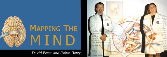
If Dr. Albert Rhoton is the foremost explorer of
human brain anatomy, then David Peace and Robin Barry are the cartographers.
For much of Rhoton’s career, Peace and Barry have been at his shoulder,
in the laboratory and the operating room, shooting pictures, making sketches
and taking notes. Back at their drawing tables and computer workstations,
they’ve taken that information and used it to draw medical illustrations
that have gained them worldwide recognition among neurosurgeons and researchers.
Peace, in his 21st year with the UF faculty, was inspired as a child by
the natural science illustrations at the Smithsonian Institute, which he
visited frequently while growing up in Washington, D.C. He dreamed of drawing
fish, reptiles, frogs and other animals for the Smithsonian, but midway
through graduate studies in medical and biological illustration at the Medical
College of Georgia in Augusta, he became interested in human anatomy and
surgery.
Barry, in her 19th year at UF, graduated with honors from Yale and earned
her Master’s of Art degree through the Johns Hopkins School of Medicine’s
renowned medical illustration program. It was there that she became fascinated
with the brain while observing surgery.
“Before you know much about the human brain, you think about it as
a gelatinous mass,” Barry says. “Then, the more you delve into
it, the more you realize how much you don’t know. And gradually you
realize it’s a never-ending odyssey of learning.”
Peace says the question he hears most often from people curious about his
profession is “Why do they need medical illustrators when you have
cameras?”
He responds with a list of things that can be done by skilled medical illustrators
that can’t be done with cameras — like erasing blood and other
elements in the surgical field that obscure the anatomy of a certain brain
region.
“We can give a three-dimensional appearance to a lot of our illustrations,
and draw in something that is invisible to the camera’s eye —
like a beam of radiation. We can go places conceptually that a camera cannot
go,” he adds.
New computer technology also gives illustrators unprecedented capabilities
to quickly enhance pencil sketches and photos with color, highlights and
complementary backgrounds, and remove distracting elements, Peace says.
Both Barry and Peace have embraced computers to achieve their artistic goals
more quickly and effectively. Barry says much of the photo/illustration
refinements she used to do by hand with paint brushes, air brushes and colored
pencils are now done in sophisticated illustration software.
Her latest challenge involves using software that merges matched pairs of
color slides into a single image that, when projected and viewed through
stereo glasses, appears three-dimensional. The technique, used in several
popular magazines to lend drama to pictures, has a more serious purpose
in neurosurgery — to add visual depth to images of brain structures
to be operated on.
Peace also has ventured into the computer world as designer/manager of the
World Wide Web site for UF’s Department of Neurosurgery (www.neurosurgery.ufl.edu).
Barry and Peace also say the chance to meet and work with leading neurosurgeons
from around the world who come to UF for advanced training is a great career
bonus.
“Some of the foreign surgeons have told me they have kept the reprints
(of my illustrations) in the operating room while performing surgery,”
said Peace. “That is especially gratifying.”